Breast cancer remains one of the most prevalent health concerns for women globally, prompting extensive research into effective diagnostic tools. While the standard for breast cancer screening is mammography, advancements in imaging technology have led to the exploration of other modalities, such as chest computed tomography (CT) scans. Though not routinely employed for breast cancer diagnosis, CT scans can sometimes reveal unsuspected findings that warrant further investigation.
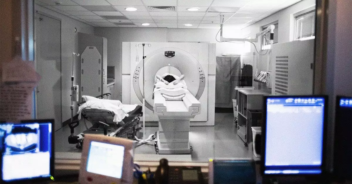
A chest CT scan uses X-ray technology to create comprehensive, cross-sectional images of the body. By acquiring multiple 2D images, healthcare professionals can generate a detailed 3D reconstruction of the internal structures, aiding in the diagnosis of various conditions. Typically, CT scans are more prevalent for assessing injuries, diseases, or conditions affecting the lungs and abdomen. They utilize contrast dyes, which can enhance the visibility of soft tissues, blood vessels, and organs, thus providing a clearer picture for medical evaluation.
In certain instances, incidental breast findings may emerge during a CT scan aimed at diagnosing non-breast-related conditions. Such incidentalomas, as they are often called, could potentially indicate cancer. Research indicates that a significant 28% of incidental findings in CT scans may reveal breast lesions, suggesting that breast abnormalities can sometimes be picked up inadvertently during other diagnostic imaging procedures.
Although CT scans are not a standard diagnostic tool for breast cancer, they do have specific applications in the clinical setting. In particular, healthcare providers may use a chest CT to determine if breast cancer has metastasized to other organs, including the lungs or liver. Furthermore, a CT-guided needle biopsy may be administered where a CT scan helps direct a biopsy needle to specific breast lesions, allowing for accurate tissue sample collection for laboratory analysis.
However, it is essential to underscore that mammograms remain the gold standard for breast cancer screening. Mammography employs low-dose X-rays specifically designed to capture images of breast tissue with reduced radiation exposure. This standard tool serves both diagnostic and screening purposes, enabling healthcare providers to detect unusual changes, masses, or other indicators of potential malignancies effectively.
Aside from mammography and CT scans, several other imaging modalities may be utilized in breast cancer evaluations. Breast ultrasounds, for example, leverage sound waves to create detailed images of breast tissue and can be particularly effective in cases where mammograms show anomalies. Similarly, breast MRI scans utilize magnetic fields and radio waves for enhanced imaging, allowing for better visualization of complex breast structures.
For any diagnostic procedure, thorough evaluations are critical. Factors such as age, family history, and personal medical history all play a role in determining the most suitable screening method. It’s not uncommon for patients to undergo a combination of imaging tests to achieve the most accurate diagnosis.
Awareness of breast cancer symptoms is crucial for early diagnosis. Individuals should be vigilant for any changes in breast tissue, including lumps, swelling, or alterations in the skin texture. Unusually shaped masses or changes in nipple appearance could signal underlying issues necessitating immediate medical attention.
Routine screenings are vital since they provide opportunities for early detection, which is paramount in improving treatment outcomes. Studies affirm that when breast cancer is found in its early stages, the chances of successful treatment and long-term survival significantly increase.
A 2023 study reported alarming statistics indicating that radiologists missed over 64% of incidental breast cancer findings on chest CT scans. This underlines the necessity for healthcare providers to scrutinize such scans meticulously, especially as these imaging tests frequently reveal breast tissue. Clear signs of possible malignancies that may be detected on a CT scan include irregular-shaped masses or lesions that exhibit rim enhancement with contrast dye.
While CT scans are not the primary method for breast cancer diagnosis, they can occasionally reveal valuable incidental findings. For optimal breast cancer detection and patient outcomes, it is essential to balance various imaging modalities and stay proactive about screening recommendations. As breast cancer remains a significant health issue, a comprehensive approach involving multiple diagnostic tools can lead to earlier detection and more effective treatment strategies. Individuals should always consult with their healthcare professionals regarding any unusual changes in their breast health and adhere to screening guidelines to facilitate early intervention.

