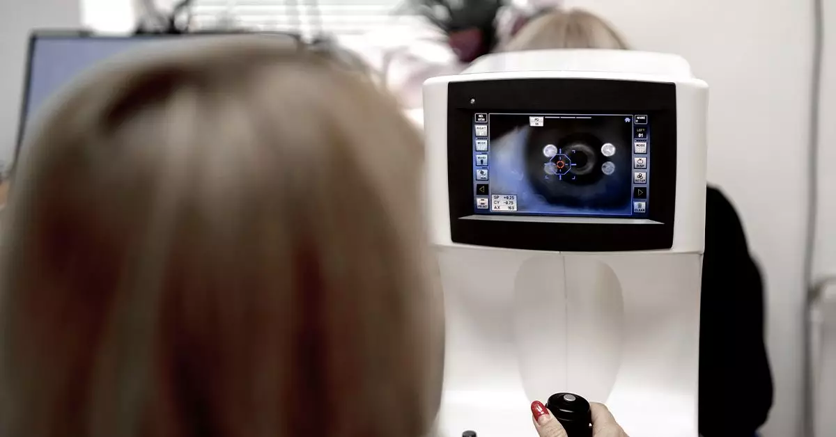
Diabetic retinopathy is a significant concern for individuals living with diabetes, as it poses a risk of severe vision impairment and blindness. Fundus photography has emerged as a crucial technique in the early detection and management of this condition. This article delves into the intricacies of fundus photography, its application in diagnosing diabetic retinopathy, and highlights its unique innovation in the realm of eye health.
Fundus photography involves capturing detailed images of the interior surface of the eye, particularly the retina, allowing healthcare providers to examine it for any signs of disease. During the procedure, a specialized fundus camera is utilized, which employs a flash of light to illuminate the retina while taking high-resolution images. While traditional fundus photography is performed in clinical settings, recent advancements have made it possible for individuals to leverage smartphone technology to capture these vital images.
This method, commonly referred to as Selfie Fundus Imaging (SFI), allows patients to take their own pictures of the retina using a smartphone after applying pupil-dilating eye drops. A 2022 study showed that SFI could produce images comparable to those captured with professional equipment, enhancing accessibility to screening for diabetic retinopathy, particularly in underserved populations.
The identification of diabetic retinopathy through fundus photography involves recognizing distinct abnormalities in the retinal images. Some of the critical indicators include:
1. **Microaneurysms**: Often the earliest sign of diabetic retinopathy, these small red spots signify the presence of fragile blood vessels, marking the onset of potential eye deterioration.
2. **Hemorrhages**: These appear as dark red patches in various shapes, indicating bleeding in the retina due to ruptured capillaries, which results from weakened vascular structures.
3. **Hard Exudates**: These yellowish-white lesions suggest lipid deposits that have leaked from blood vessels, providing insights into metabolic disparities affecting the eye.
4. **Cotton Wool Spots**: Representing localized edema due to a lack of blood supply, these fluffy white patches are an indicator of possible ischemic changes in retinal tissue.
5. **Neovascularization**: The formation of new, abnormal blood vessels indicates a progression to more severe stages of diabetic retinopathy and necessitates immediate medical intervention.
Understanding these indicators empowers both patients and healthcare providers to engage in timely and effective management of diabetic retinopathy.
For individuals diagnosed with diabetes, the importance of regular eye examinations cannot be overstated. The American Diabetes Association recommends that those aged 10 and older with diabetes should undergo comprehensive eye exams at least once a year. Early detection through fundus photography not only allows for timely intervention but also significantly reduces the risk of vision loss. It’s crucial for individuals, especially those who are expecting or have been diagnosed with gestational diabetes, to prioritize dilated eye exams to monitor potential changes.
During these examinations, patients may experience temporary side effects, such as blurry vision and heightened sensitivity to light due to the dilating eye drops. Thus, it is advisable to refrain from driving or engaging in activities that require clear vision immediately after the procedure.
Regular fundus imaging serves as both a preventative and a diagnostic tool within the wider context of diabetes management. However, patients must also adopt comprehensive strategies for managing their diabetes, which include maintaining a balanced diet, engaging in regular physical activity, and adhering to prescribed medication regimens. Such practices not only support eye health but also contribute to overall well-being, significantly helping to delay or prevent the onset of diabetic retinopathy.
Moreover, education on recognizing early symptoms such as fluctuating vision, the appearance of floaters, or any sudden changes in visual acuity is vital for proactive health management. Promptly reporting these symptoms to healthcare providers can enhance chances of preserving vision.
Fundus photography represents a pivotal advancement in the realm of diabetic eye care, providing essential insights that can avert significant visual impairment stemming from diabetic retinopathy. The integration of smartphone technology through Selfie Fundus Imaging offers a new frontier in accessibility, allowing individuals to take an active role in their health management. As research continues to evolve,, it is imperative for those at risk of diabetic retinopathy to remain vigilant about their eye health, seeking regular screenings and maintaining their overall well-being to ensure a brighter future for their vision.

